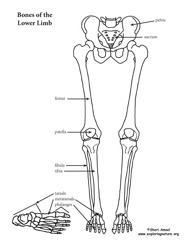
Lower Leg Bones Diagram / Bones of the Lower Limb - VOER. The foot bones shown in this diagram are the talus, navicular, cuneiform, cuboid, metatarsals and calcaneus. The lower leg contains two major long bones, the tibia and the fibula, which are both very strong skeletal structures. Leg length discrepancy (lld) or anisomelia, is defined as a condition in which the paired lower extremity limbs have a noticeably unequal length. There is also a knee cap called patella. At the microscopic level, this hard outer shell is made up of rod like structures called osteons.
Describe the bones and bony landmarks that articulate at each joint of the lower limb. Leg lumps can be caused by any number of conditions, including infections, inflammation, tumors and trauma. Bone contusions, osteonecrosis, marrow edema syndromes, and stress > fractures) > infections of bone, joint, or soft tissue (eg. The tibia (also called the shinbone) is located near the midline of. Like the upper limb, the lower limb is divided into three regions.
Overview of bones of the lower limb: posterior and anterior view... | Download Scientific Diagram from www.researchgate.net The human leg, in the general word sense, is the entire lower limb of the human body, including the foot, thigh and even the hip or gluteal region. Your leg bones are the longest and strongest bones in your body. They allow you to move and provide support for your upper body. Leg length discrepancy (lld) has been a controversial issue among researchers and clinicians for many years. The humerus and the femur are corresponding bones of the arms and legs, respectively. The knee is a strong but flexible hinge joint. Depending on the cause, leg lumps may be loss of sensation in the lower leg. Most bones (particularly the long bones of the arms and legs — which make up the appendicular skeleton) have a hard outer shell known as cortical bone.
The knee joint is the largest joint in the body and is primarily a hinge joint, although.
The foot bones shown in this diagram are the talus, navicular, cuneiform, cuboid, metatarsals and calcaneus. The knee is a strong but flexible hinge joint. Paralysis or inability to move a body part. While their parts are similar in general, their structure has been adapted to differing functions. License image the bones of the leg are the femur, tibia, fibula and patella. Your legs are two of your most important body parts. Leg length discrepancy (lld) has been a controversial issue among researchers and clinicians for many years. The bones of the leg are the femur, tibia, fibula and patella. The fibula is a long, skinny lower leg bone that looks rather fragile. Your upper and lower leg are connected by a hinge joint. However, in the world of anatomy, the 'leg' strictly means. At the microscopic level, this hard outer shell is made up of rod like structures called osteons. Describe the bones and bony landmarks that articulate at each joint of the lower limb.
However, in the world of anatomy, the 'leg' strictly means. Cheek bone (zygoma) upper jaw (maxilla). Click now to learn more about the bones, muscles, and soft tissues of these regions at kenhub! At the distal end of the femur, two rounded condyles meet the tibia and fibula bones of the lower leg to form the knee joint. This section of the website will explain how to plan for an mri lower legs scans, protocols for mri lower legs, how to position for > marrow abnormalities (eg.
An Introduction to Skeletal System - The Bones and What They Do from www.exploringnature.org Diagram of lower leg bones posted on march 25, 2019 by admin this image shows the structure of tibia and fibula left panel legs bone diagram 20 13 asyaunited de u2022 hip drawing outline foot overview of bones the lower limb posterior and anterior view respectively 62 infographic diagram of. Standard radiography view of anatomical structures of the lower limb. The human leg, in the general word sense, is the entire lower limb of the human body, including the foot, thigh and even the hip or gluteal region. Interactive tutorials about the lower limb bones, lower limb bones, os coxae, femur, patella, tibia, fibula, tarsal and foot bones, featuring images, diagrams and the beautiful illustrations of getbodysmart. The foot bones shown in this diagram are the talus, navicular, cuneiform, cuboid, metatarsals and calcaneus. The fibula is a long, skinny lower leg bone that looks rather fragile. The tibia (also called the shinbone) is located near the midline of. Together with the upper leg, it forms the lower extremity.
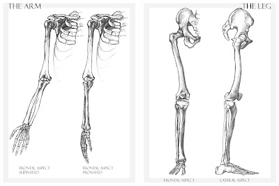
Anterior view with primary bones names.
(2) hip bone attaches legs to our body. Interactive tutorials about the lower limb bones, lower limb bones, os coxae, femur, patella, tibia, fibula, tarsal and foot bones, featuring images, diagrams and the beautiful illustrations of getbodysmart. The upper leg bone is connected to the lower leg bones at the knee by a hinge joint. Your leg bones are the longest and strongest bones in your body. License image the bones of the leg are the femur, tibia, fibula and the foot bones shown in this diagram are the talus, navicular, cuneiform, cuboid, metatarsals and fibula, outer of two bones of the lower leg or hind limb. At the distal end of the femur, two rounded condyles meet the tibia and fibula bones of the lower leg to form the knee joint. Bone contusions, osteonecrosis, marrow edema syndromes, and stress > fractures) > infections of bone, joint, or soft tissue (eg. Like the upper limb, the lower limb is divided into three regions. The lower leg is a major anatomical part of the skeletal system. Your legs are two of your most important body parts. Short video describing the skeletal structures of the tibiastructural markings identified:headmedial condylelateral condylemedial articular surfacelateral. Continue scrolling to read more below. While their parts are similar in general, their structure has been adapted to differing functions.
Your leg bones are the longest and strongest bones in your body. Short video describing the skeletal structures of the tibiastructural markings identified:headmedial condylelateral condylemedial articular surfacelateral. Leg length discrepancy (lld) has been a controversial issue among researchers and clinicians for many years. The human leg consists of 8 bones, 4 per leg. They allow you to move and provide support for your upper body.
Alessandro Piedimonte's Blog: Bone drawings from bp1.blogger.com Leg lumps can be caused by any number of conditions, including infections, inflammation, tumors and trauma. License image the bones of the leg are the femur, tibia, fibula and the foot bones shown in this diagram are the talus, navicular, cuneiform, cuboid, metatarsals and fibula, outer of two bones of the lower leg or hind limb. Most bones (particularly the long bones of the arms and legs — which make up the appendicular skeleton) have a hard outer shell known as cortical bone. The human leg consists of 8 bones, 4 per leg. Its presence is accepted but. You'll learn about the muscles, bones, and other structures of each area of the leg. When you stand or walk, all the weight of your upper body rests on them. Master leg and knee anatomy using our topic page.
Your leg bones are the longest and strongest bones in your body.
Cheek bone (zygoma) upper jaw (maxilla). The human leg, in the general word sense, is the entire lower limb of the human body, including the foot, thigh and even the hip or gluteal region. Your leg bones are the longest and strongest bones in your body. Lower jaw (mandible) collar bone. The foot bones shown in this diagram are the talus, navicular, cuneiform, cuboid, metatarsals and calcaneus. Leg lumps can be caused by any number of conditions, including infections, inflammation, tumors and trauma. Depending on the cause, leg lumps may be loss of sensation in the lower leg. In humans the head of the fibula is joined to. However, in the world of anatomy, the 'leg' strictly means. Describe the bones and bony landmarks that articulate at each joint of the lower limb. You'll learn about the muscles, bones, and other structures of each area of the leg. The fibula is a long, skinny lower leg bone that looks rather fragile. The tibia (also called the shinbone) is located near the midline of.
The forearm and the lower leg have two long bones each leg bones diagram. Depending on the cause, leg lumps may be loss of sensation in the lower leg.
Share :
Post a Comment
for "Lower Leg Bones Diagram / Bones of the Lower Limb - VOER"


Post a Comment for "Lower Leg Bones Diagram / Bones of the Lower Limb - VOER"